
3d Illustration of Human Neck Muscle Anatomy for the Education Stock Biology Diagrams Learn everything about the neck anatomy with this topic page. Click now to study the muscles, glands and organs of the neck at Kenhub!

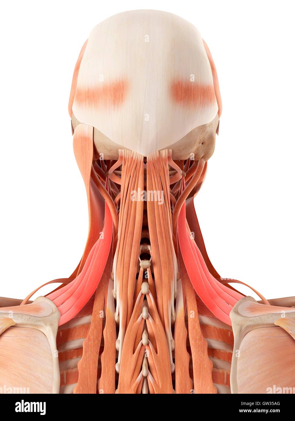
The muscles of the neck are present in four main groups. The suboccipital muscles act to rotate the head and extend the neck. Rectus capitis posterior major and Rectus capitis posterior minor attach the inferior nuchal line of the occiput to the C2 and C1 vertebrae respectively.
The Muscles of the Head and Neck: 3D Anatomy Model Biology Diagrams
3D interactive tutorials on the anatomy of the neck, including the anatomical organisation, musculature, larynx, pharynx, blood supply and innervation.

Your neck muscles support your head and help you do a range of movements. They also assist with chewing, swallowing and breathing. Muscles of the Neck: Neck Anatomy Muscles Pictures There are many muscles around the neck that help to support the cervical spine and allow you to move your head in different directions. Here is a list of the many muscles that exist in the neck. Longus Colli & Capitis - Responsible for flexion of the head and neck.
Neck Anatomy Pictures Bones, Muscles, Nerves Biology Diagrams
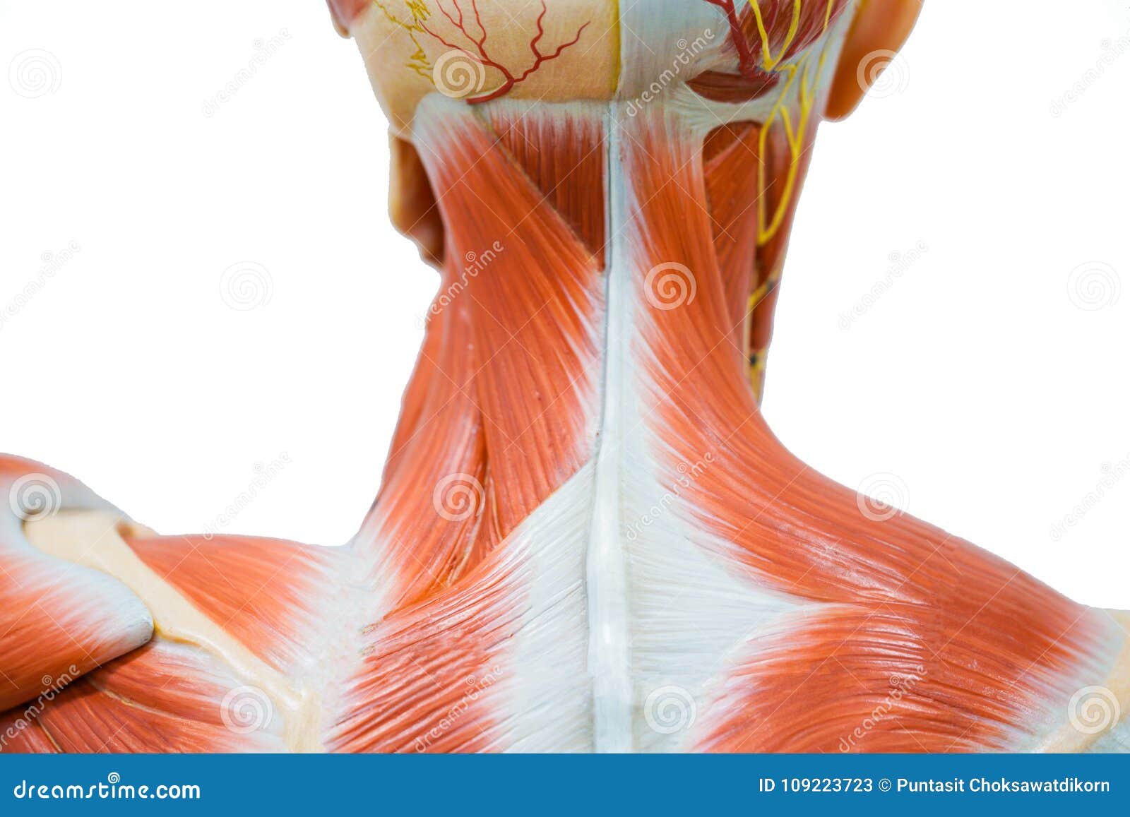
The neck is a body part that connects the head with the rest of the body. It contains various organs and is built of different tissue, including many skeletal muscles that provide movements of the head. These muscles are called the neck muscles. The main function of these muscles is to permit movements of the neck and head. Also, they provide structural support for the head. The muscles of the Your neck muscles allow you to turn your head from side to side, forwards, and backwards. Learn which muscle groups get tight and restricted. This comprehensive guide details the anatomy of the neck, including the cervical spine, larynx, thyroid & lymphatics. Learn more about human anatomy here.
