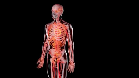
1 Left figure shows an already simplified representation of the human Biology Diagrams The intercostal arteries are a pair of arteries on either side of the body that send blood to various areas of the torso, including the vertebrae, spinal cord, back muscles, and skin. Superior

The aorta, the largest artery in our body, exemplifies this type. - Muscular Arteries: Found farther from the heart, these arteries contain more smooth muscle tissue and fewer elastic fibers. They regulate blood flow to specific organs and regions. ### 2. Artery Wall Layers. The walls of arteries consist of three distinct layers:

Major Arteries of the Body: The Aorta, Head, Neck & Torso Biology Diagrams
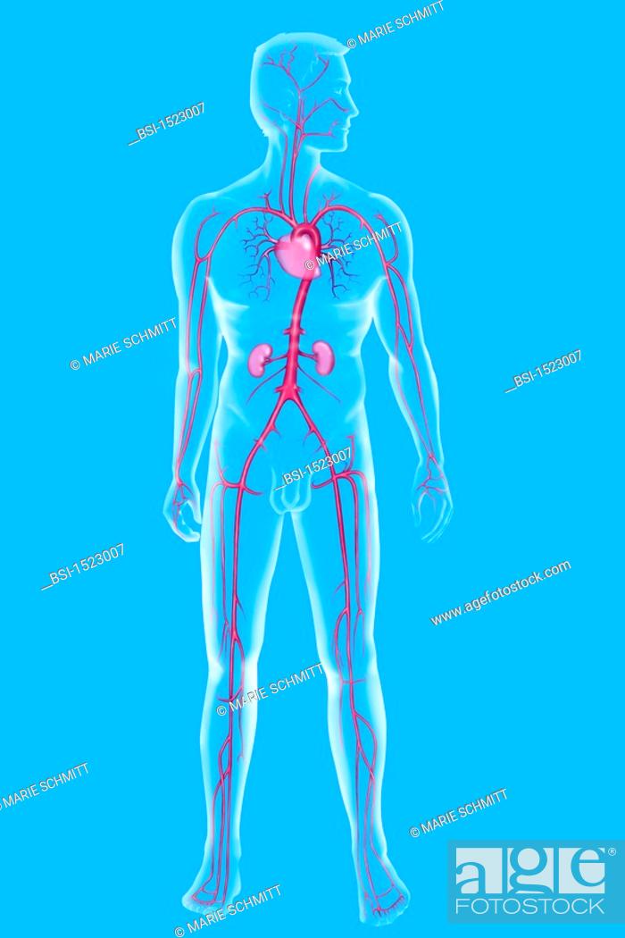
Arteries make up a major part of the circulatory system, with the veins and heart being the other main components. Arteries make up tubelike structures responsible for transporting fluid (i.e., blood for the circulatory system and lymph for the lymphatic system) to and from every organ in the body. Mainly, arteries manage the transportation of oxygen, nutrients, and hormones through our bodies

As arteries extend further from the heart, they usually become narrower. Additionally, arteries will continually branch off into smaller arteries, arterioles and capillaries, forming the vascular bed. The major arteries in the body are: The aorta. The largest artery in the body, which connects directly to the left ventricle of the heart.
Anatomy, Arteries Biology Diagrams
Your circulatory system, or cardiovascular system, supplies oxygen and nutrients to your whole body and removes waste through your blood. Your heart pumps blood that flows through your arteries, veins and capillaries. These blood vessels and your heart form your circulatory system. They work together to ensure your cells have what they need.